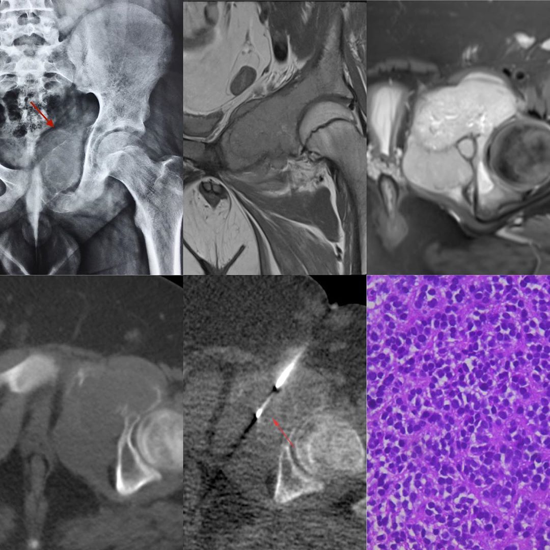
This is a 13-years old boy with pain in the left hip and swelling on palpation.
The radiograph shows an expansile osteolytic lesion with abnormal soft tissue involving the left iliopubic ramus. The T1W coronal and PDFS axial MRI images show the lesion well. The CT scan shows that the cortex is markedly thinned, making the biopsy with an 18G coaxial biopsy gun very easy and straightforward.
The histopathology was a classic round cell tumor consistent with Ewing sarcoma.
#CTbiopsy #Ewingsarcoma
Ewing Sarcoma Acetabulum
Blogs
Latest From Gallery


