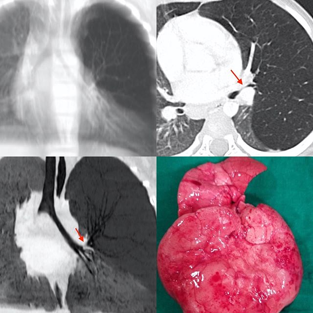
This 3 years old boy came with sudden breathlessness. A simulated radiograph from the CT scan images shows increased lucency of the left upper and mid-zones, which on the axial CT scan is seen a left upper lobe overinflation due to a high-grade stenosis of the left upper lobe bronchus (arrow). The minIP image shows this well (arrow). The operated overinflated lung is seen in the last panel.
Congenital Lobar Emphysema (CLE)

Blogs
Categories
Tags
The REF started in fun! In August 1996, after the Saturday radiology meeting in KEM, a few residents and Ravi and Bhavin were sitting at the “katta” having “chai”, when the usual subject of resident education came up.
Contact Us
91 22 6617 3333
info@refindia.net
"F", 1st Flr, Bhaveshwar Vihar, 383, S.V.P Road, Mumbai- 400004, India.


