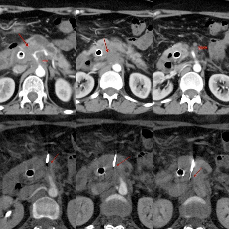
This 56-years old lady presented with a pancreatic head mass. She was known to have Hodgkin’s lymphoma in the past, and a biopsy was needed for the exact histology.
The mass encases the celiac axis and the portal vein and we can see the CBD stent that was put in to relieve the obstructive jaundice. The pancreatic duct is also dilated.
There was a good window between the celiac axis and the SMA towards the right. I gave intravenous contrast to ensure that I could visualize the vessels and accurately place the canula away from all the vessels and get adequate tissue.
The diagnosis was adenocarcinoma.
Rule: Don’t be afraid to use intravenous contrast to delineate vessels, when they crowd the target lesion.


