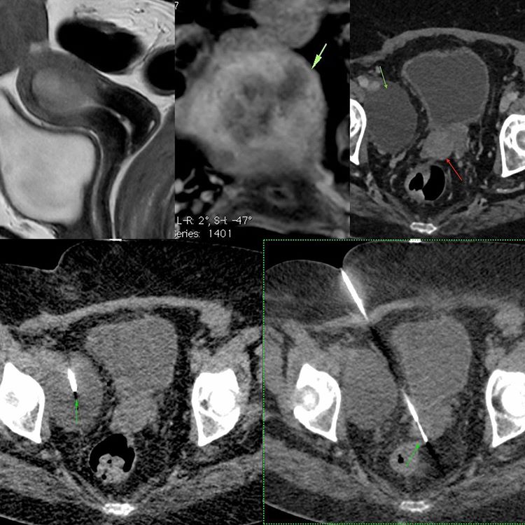
This is an 82-years old lady who in September last year had an MRI of the pelvis showing endometrial carcinoma with myometrial involvement, but without extra-uterine spread or nodes. The T2W sagittal shows the lesion well while the post-contrast T1W coronal shows the focal myometrial invasion well.
She underwent hysterectomy with salpingo-oophorectomy. She came back for a repeat CT scan in April 2020, which showed a focal nodule in the surgical bed (red arrow) and a lymphocele (green arrow) in the right iliac bifurcation region.
I did a biopsy of the focal nodule in the surgical bed using a 20 cm long 20G coaxial biopsy gun. The nodule was hard and fibrotic and it seemed as if this was just post-operative fibrosis. The surgeon also wanted an aspiration of the lymphocele, which was also done.
The diagnosis turned out to be adenocarcinoma recurrence. The lymphocele showed straw-colored fluid consistent with lymphocele.


