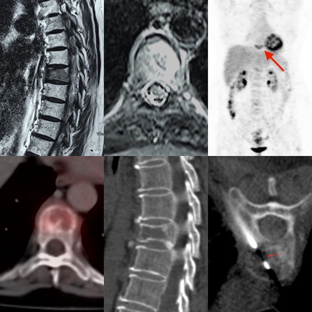
This is a 53-years old with backache. An MRI was done, which showed a focal lesion in the D9 vertebral body (T2 sagittal and STIR axial). Somehow she landed up directly with a hematologist, who asked for a PET/CT. This was the only lesion showing mild activity.
A lesion such as this is indeterminate for etiology. There is no clue to the diagnosis on the MRI itself. Hence statistically, this would be metastasis, plasmacytoma or lymphoma unless proven otherwise and in our country, tuberculosis also needs to be kept in mind.
The CT scan obtained at the time of the biopsy showed the lesion particularly well (sagittal image). The biopsy was performed using a standard costo-vertebral approach from he left under sedation with an 18G coaxial biopsy gun. It turned out to plasmacytoma.
#CTbiopsy #plasmayctoma #spinetumor


