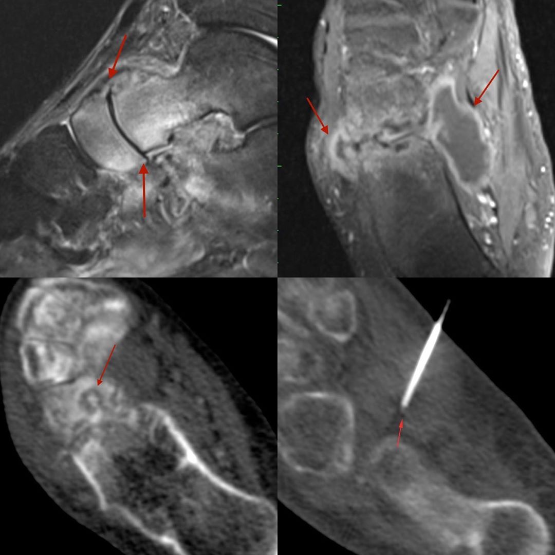
This is a 52-years old man with ankle and foot swelling for 2 years. He had an obvious mid-foot arthopathy, but no diagnosis had been established.
An MRI showed both talonavicular (STIR sagittal) and calcaneocuboid (T1W post Gd axial) involvement. A CT scan at the time of the biopsy showed a cuboid osteolytic lesion with a sequestrum.
The biopsy needle was advanced into the collection seen on the MRI and yielded frank pus, which was MTB positive on PCR and culture. A bone biopsy was obviated.
However, if no frank pus is found in such procedures, than biopsy through the sequestrum and any other osteolytic area becomes essential.
#CTbiopsy #tuberculosisfoot #tuberculosis


