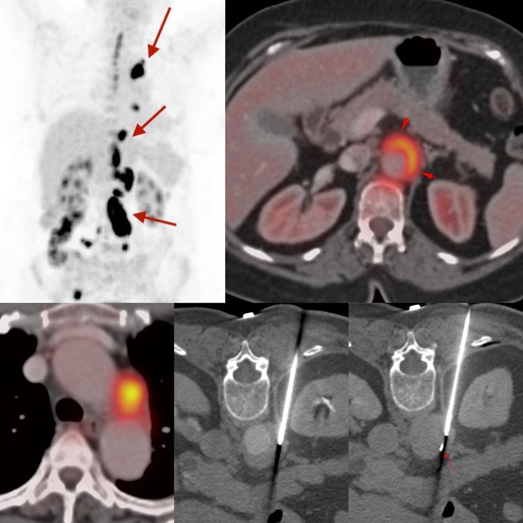
This is a 67-years old lady with fever, who presented with fever. A CT scan showed peri-aortic soft tissue. A PET/CT showed non-contiguous lesions, adjacent to the aortic arch and in relation to the abdominal aorta.
A biopsy was needed to differentiate peri-aortitis from IgG4 disease from lymphoma.
The biopsy was done using a 20G coaxial biopsy gun with the use of intravenous contrast to delineate the aortic lumen. Basically as long as we don’t biopsy the aortic intima, these biopsies are quite straightforward, though in practice, few radiologists actually attempt to do them.
The diagnosis was inflammation, confirming that this was idiopathic peri-aortitis. There was no tumor or IgG4 disease. The lady did well on treatment.


