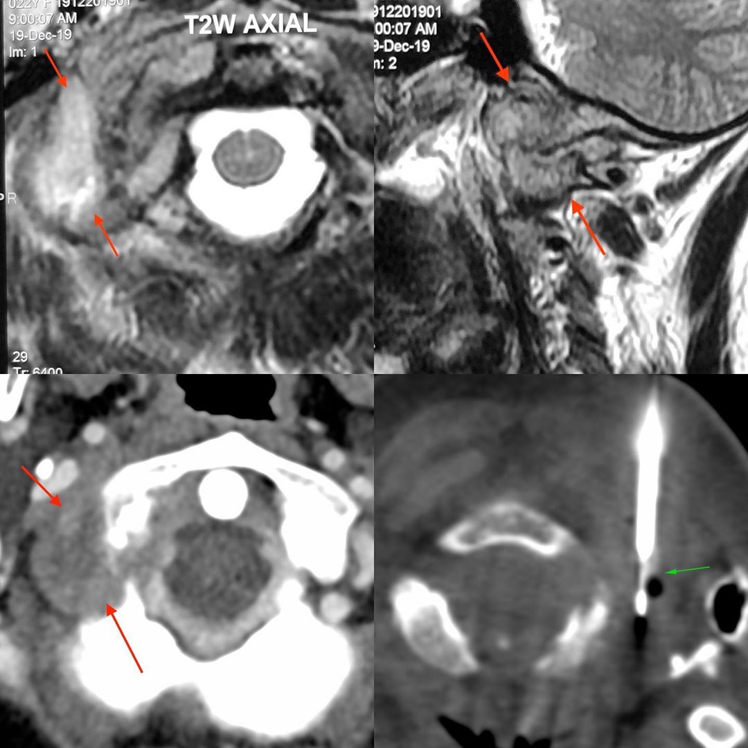
This is a 22-years old lady who presented with neck pain with painful movements. An MRI showed an osteolytic lesion involving the right occipital condyle (axial T2) and the adjacent occipito-atlantal joint (sagittal T2). She was sent for a biopsy.
We did a contrast CT to understand the anatomy of the lesion and found that there was easy access with the vertebral artery inferior to the lesion and not in the path of the needle. Frank pus was aspirated that was PCR positive for MTB.
#tuberculosis #craniovertebraljunction #CVJ #CTbiopsy
Occipital Condylar TB – One More Unusual Site
Blogs
Latest From Gallery


