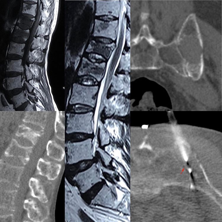
This is a 60-years old man with severe backache. The MRI (T1W and T2W sagittal images) showed multiple focal lesions. The CT scan at the time of biopsy (sagittal image) showed multiple lesions and the plan was to target the anterior D12 vertebral body lesion. This biopsy had a certain degree of difficulty and there was no guarantee we would have got adequate material for a diagnosis.
Since there were multiple lesions, I asked the technologist to run a pelvic scan, which showed large left sacral and iliac bone lesions. After that, it was the easiest thing to put an 18G coaxial core biopsy gun into the iliac bone lesion to get the diagnosis of a plasma cell neoplasm, which given the multiple lesions was suggestive of multiple myeloma.


