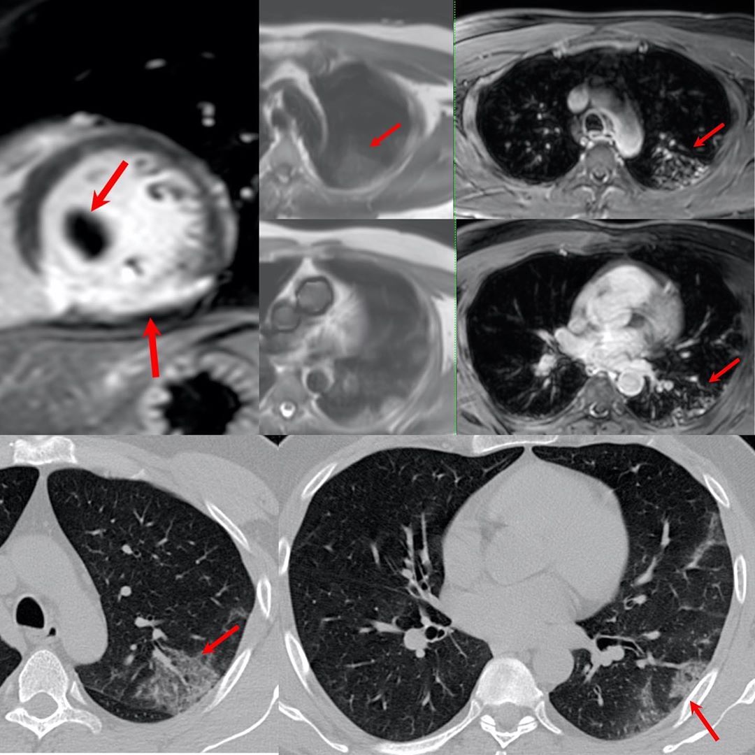
This 53-years old man came with a recent inferior wall infarct, for viability imaging.
The top left image shows the inferior wall infarct with a thrombus (arrow). The HASTE and contrast VIBE MRI images showed wedge-shaped areas (arrows) of altered signal in the posterior segment of the left upper lobe and the superior segment of the left lower lobe of the lung.
A CT scan done immediately thereafter shows typical COVID-19 lung changes (arrows) in the same segments and the diagnosis was confirmed later with RT-PCR testing.


