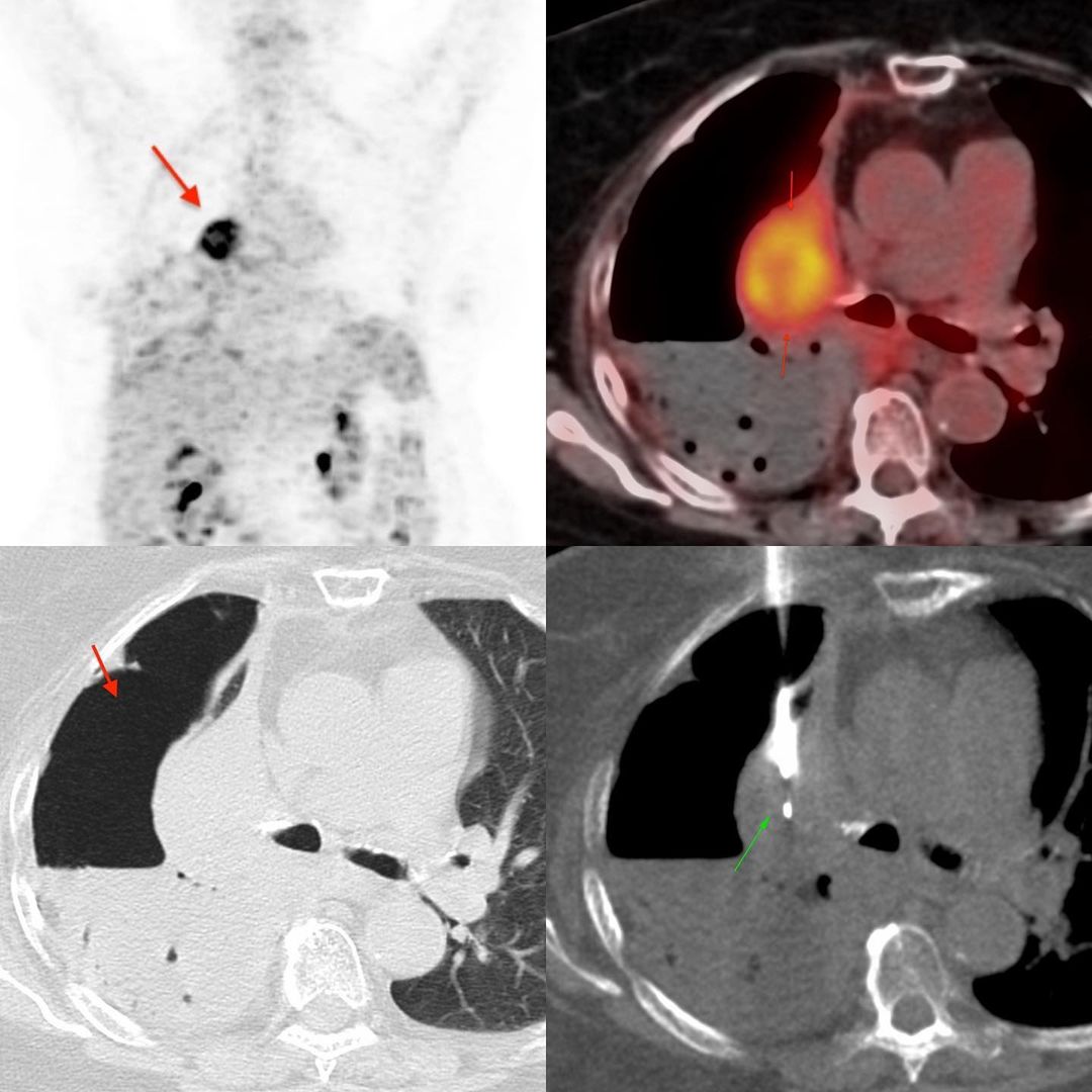
This 81 years old lady presented with a hydropneumothorax. A CT scan showed a possible mass in the atelectatic right upper lobe. A PET/CT confirmed a mass with uptake.
She came with a chest tube for a CT guided biopsy, which made the biopsy very simple and straightforward. The biopsy, using an 18G coaxial biopsy gun was uneventful. Enough material could be obtained for all the mutation studies, which included EGFR, ALK, ROS1, PDL1.
Apart from hemorrhage, the only other complication to worry about is a pneumothorax. When it is already present and the patient has a chest tube, then even that is no longer an issue and we can go through the pneumothorax or as in this case, the hydronpneumothorax and safely biopsy the lung lesion.


