This 29-years old lady presented with ankle pain.
The frontal radiograph shows a well-defined osteolytic lesion with a narrow zone of transition. The sagittal CT scan shows the same finding. The MRI shows a typical T2 dark lesion with fluid levels, findings that, in the setting of a benign lesion, are suggestive of a giant cell tumor (GCT) with a secondary aneurysmal bone cyst (ABC) component.
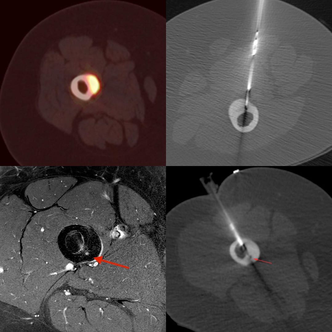
The question was how to biopsy. For bone tumors, the biopsy should be done along the plane of the expected surgical incision. I drew these lines on the axial CT and sent them to the orthopedic oncosurgeon, who asked me to biopsy along the red line, i.e an anteromedial approach.
The final diagnosis was giant cell tumor (GCT). #CTbiopsy #bonetumor #benignbonetumor #giantcelltumor


