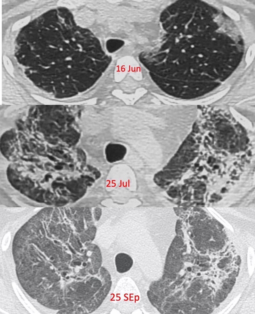
This 47-years old had COVID-19 in mid-June. Her CT scan of 16 Jun shows typical subpleural areas of ground glass. In most patients, these resolve completely or with residual subtle ground glass or reticular opacities, as we have been seeing. This lady had particularly severe disease and the scan of 25 Jul shows traction bronchiectasis and distortion of architecture with volume loss, findings that are suggestive of fibrosis. Her repeat scan of 25 Sep shows further volume loss with definite fibrosis.
This is not a common occurrence in most COVID-19 patients, but when there is honeycombing or traction bronchiectasis with architectural distortion, that is the time we can say for sure that there is post-COVID fibrosis.


