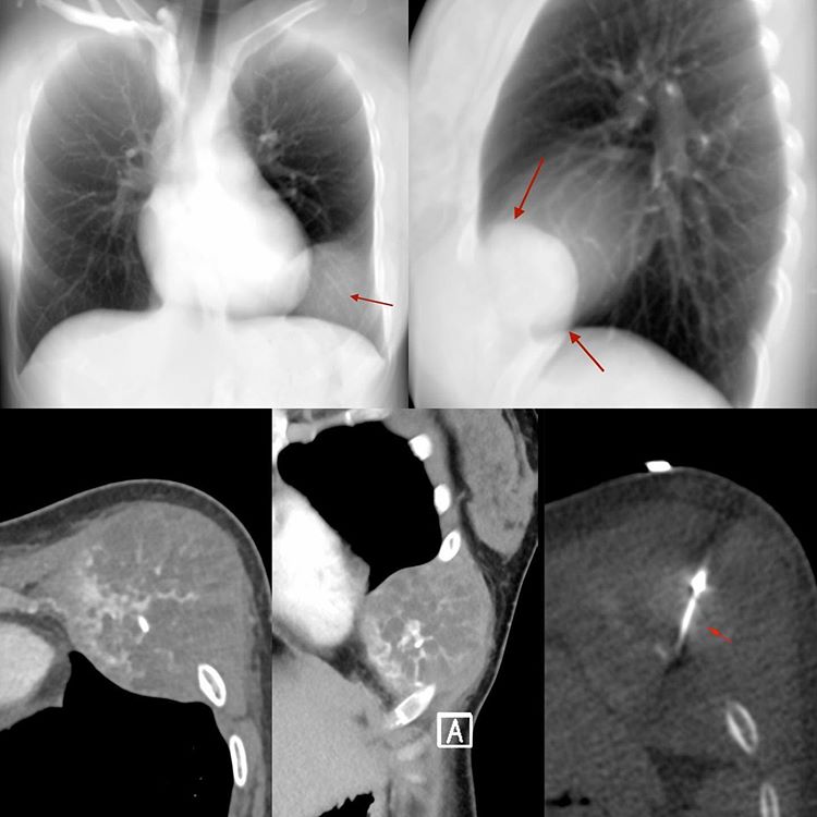
This 20-years old woman presented with a slowly growing mass over the left mid chest wall. The simulated radiographs (using the Mean function on the CT scan) show an extra pleural mass with involvement of the left 6th rib. The CT scan images in the axial and coronal planes show an enhancing mass with an osteoid matrix.
The biopsy was obviously simple performed along the plane of expected surgery.
It was a high grade osteosarcoma.


