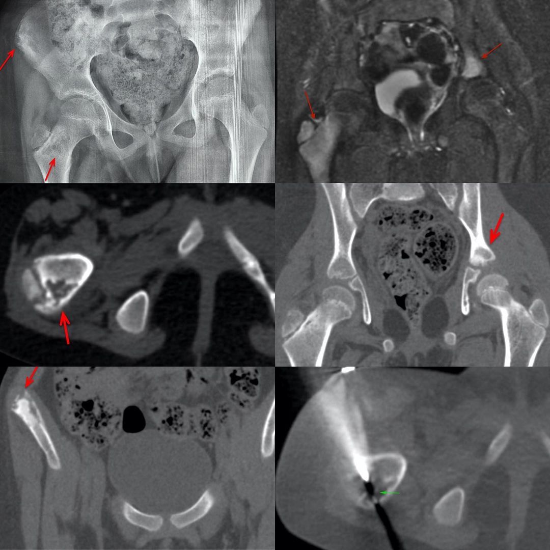
This 8-years old girl presented with pain in the right hip. The radiograph showed areas of osteolysis and sclerosis in the right femoral neck and greater trochanter and in the right iliac crest. The MRI showed another lesion in the left acetabular roof. All 3 lesions were well seen on CT scan. A CT guided biopsy of the lesion involving the right trochanteric physis and subjacent bones was negative for any definitive etiology.
Based on the multiple lesions and the clinical presentation and biopsy findings, a diagnosis of CNO was made. She delayed her treatment but eventually responded to pamidronate.


