This is a 74-years old man, smoker, who presented with fever and cough.
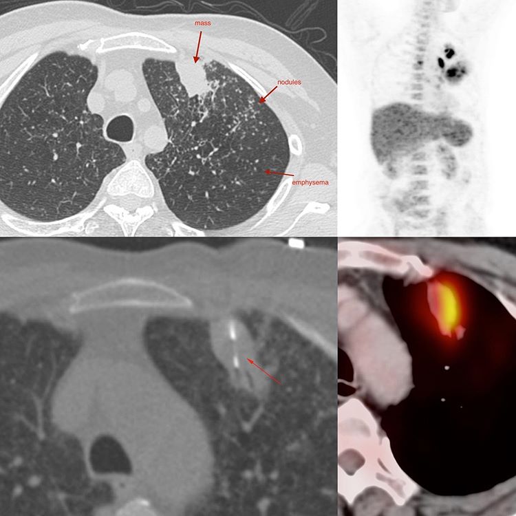
The CT scan showed a focal lesion in the anterior segment of the left upper lobe with extensive “miliary” nodules bilaterally. There was background emphysema.
The working diagnosis was malignancy with spread, but even with “miliary” metastases, they are never the classic “miliary” nodules we get with tuberculosis. The metastatic lesions invariably show some difference in size.
A PET/CT was also performed in view of the possibility of malignancy prior to the biopsy. The lesion was active, along with a few mediastinal nodes.
The biopsy was simple with a 20G coaxial biopsy gun and showed PCR, culture and histopath positive tuberculosis.
It does not mean that we think of tuberculosis and not work-up the patient. Every such patient has to have some form of histology and microbiology work-up. However, in a tuberculosis endemic country like ours, we should never be surprised when lesions that look like malignancy turn out to be tuberculosis.


